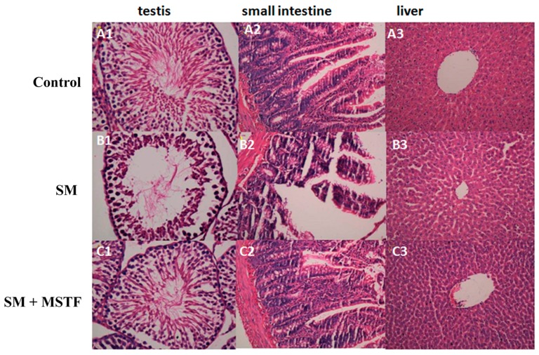
Figure 2.
Representative photographs showing the histopathologic changes in testis, small intestine and liver tissues on the third day post-exposure (n = 5 rats/group). (A1–A3) Control (normal) group; (B1–B3) SM-treated group: the decrease of sperm cells, the villus epithelial shedding of small intestinal cells, the defects of the gut mucosal barrier, as well as the swelling of liver cells; (C1–C3) SM + MSTF group treated with MSTF (120 mg/kg) 1 h after SM treatment. The damages were ameliorated by MSTF treatment (H&E, 40×).

