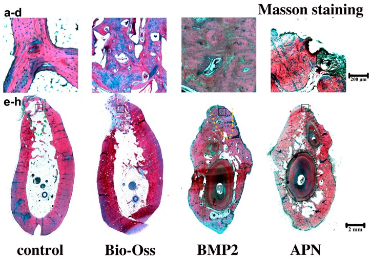
Figure 5.
Decalcified toluidine Masson staining of extraction sockets following the different 12-week treatments: (a) The magnifying control group; (b) Bio-Oss group; (c) BMP2 group; (d) APN group; (e–h) represent the integral extraction socket of control group (e); Bio-Oss group (f); BMP2 group (g); APN group (h). The black square represents the magnifying area. The dashed line represents the extraction sockets. New new bone is shown in red. Magnification: upper panel 40×; lower panel 4×. n = 4 for each group.

