Figure 4.
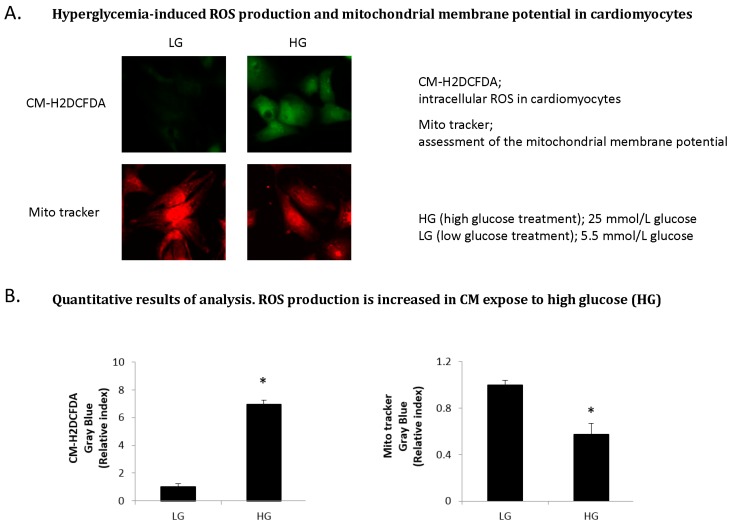
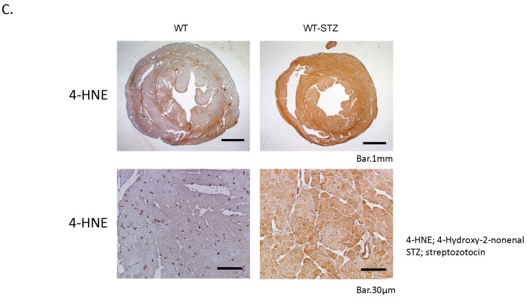
ROS production and oxidative stress in the diabetic heart in vitro and in vivo. (Copyright 2015 American Diabetes Association from [44]. Reprinted with permission from The American Diabetes Association). (A) Hyperglycemia-induced ROS production and mitochondrial membrane potential in cardiomyocytes. Intracellular ROS level is increased in cardiomyocytes (CM) expose to high glucose (HG) by using chloromethyl-2,7-dichlorodihydro-fluorescein diacetate (CM–H2DCFDA). There was loss of mitochondrial membrane potential (ΔΨm) in CM expose to high glucose as indicated by a decrease in the fluorescence intensity assessed using Mito Tracker red; (B) Quantitative results of analysis. ROS production is increased in CM expose to high glucose (HG). CM-H2DCFDA, intracellular ROS in cardiomyocytes. Mito tracker, assessment of the mitochondrial membrane potential. HG (high glucose treatment), 25 mmol/L glucose; LG (low glucose treatment), 5.5 mmol/L glucose. * p < 0.05 vs. LG. Error bars indicate s.e.m. n = 4–6; (C) Cardiac oxidative stress in the diabetic heart. Immunohistological staining (brown) of 4-hydroxy-2-nonenal (4-HNE) in the hearts of wild-type (WT), wild-STZ (WT-STZ) mice. Upper panel is 20×, lower panel is 400×; Scale bar, 1 mm and 30 μm, respectively. Cardiac 4-HNE, a major marker of oxidative stress, is up-regulated in myocardium in WT-STZ heart compared to WT heart.


