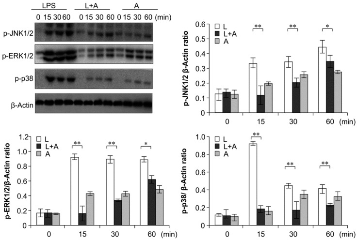
Figure 7.
Western blot analysis for inhibition of MAPK activation by humanized anti-TLR4 antibody Fab. Cells were pretreated with humanized anti-TLR4 antibody for 2 h and further incubated in presence or absence of LPS (1 µg/mL). After immunoblotting, the phosphorylation levels of ERK1/2, JNK1/2, p38 were identified using phosphor-specific antibodies. β-Actin was used to ensure equal loading. L: LPS, A: humanized anti-TLR4 antibody. Data are shown as mean ± SD (n = 3, * p < 0.05, ** p < 0.01 versus LPS group).

