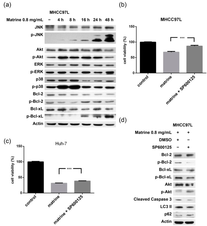
Figure 3.
Role of the JNK-Bcl-2/Bcl-xL pathway in apoptosis and autophagy. (a) MHCC97L cells were treated with matrine (0.8 mg/mL) for 4, 8, 16, 24, and 48 h. After matrine treatment, proteins in cell lysate were separated by SDS–PAGE. JNK, p-JNK, Akt, p-Akt, p38, p-p38, ERK, p-ERK, Bcl-2, p-Bcl-2, Bcl-xL, and p-Bcl-xL were detected by immunoblotting; (b), (c) and MHCC97L and Huh-7 cells were treated with matrine (0.8 mg/mL) and matrine (0.8 mg/mL) + SP600125 (90 nM) for 48 h. Cell viability was then analyzed by CCK-8 cell viability assay. *** represents a statistical significance at p < 0.001. p-Values of (b,c) were 0.0006 and 0.0003, respectively; and (d) After the separation of proteins in cell lysate with SDS–PAGE, Bcl-2, p-Bcl-2, Bcl-xL, p-Bcl-xL, p62, LC3, and the cleavage of caspase 3 were detected by immunoblotting.

