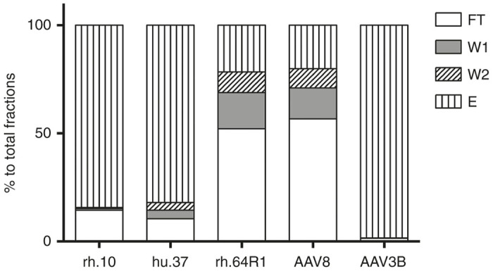
Figure 1.

Vector genome distribution among the AVB column fractions. AAV vectors were diluted in binding buffer AVB.A (for AAV3B, culture supernatant was buffer-exchanged into the binding buffer) and then loaded onto the AVB column. Fractions from flow through (FT), AVB.A wash (W1), AVB.C wash (W2), and elution (AVB.B) (E) were collected. Vector genome copies were determined by real-time PCR.
