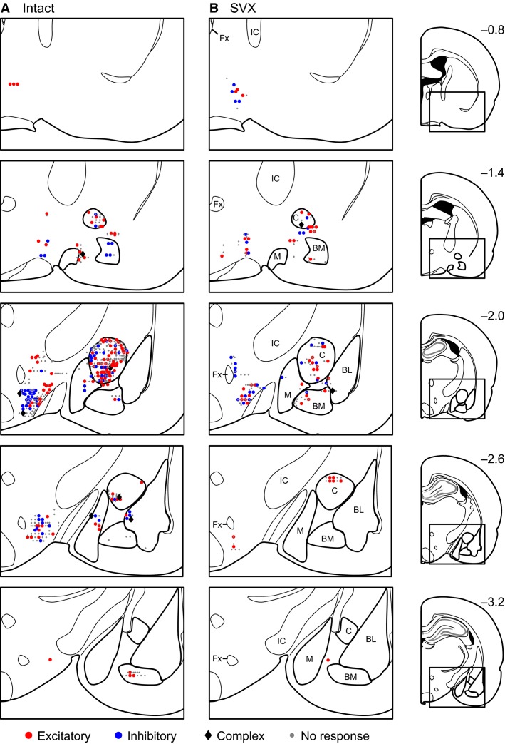
Figure 6.
Recording locations. Each symbol represents the location of excitatory (red circle), inhibitory (blue circle), or complex (black diamond) responsive neurons or nonresponsive (gray dot) neurons recorded in an intact (A) or SVX (B) rats. Each value on the left side of each section indicates the distance (mm) from bregma. BL, basolateral nucleus of the amygdala; BM, basomedial nucleus of the amygdala; M, medial nucleus of the amygdala; C, central nucleus of the amygdala; IC, internal capsule; Fx, fornix.

