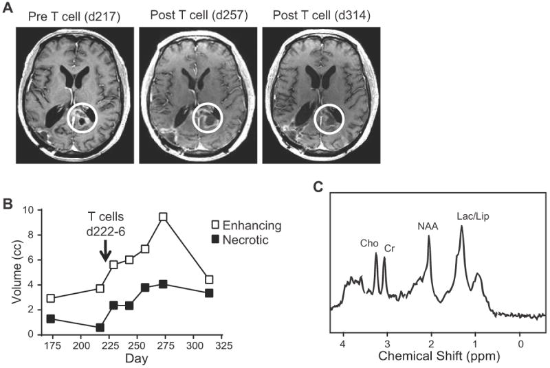
Fig. 5. MR Imaging of UPN033 shows increased necrotic tumor volume following administration of IL13-zetakine+ CD8+ T cell clones.

A, T1-weighted post-gadolinium MRI before and after the 5 daily infusions of autologous IL13 (E13Y)-zetakine+/HyTK+ CD8+ CTL. B, MRS analysis of enhancing volume (indicative of neuroinflammation) and necrotic volume at the recurrent site following re-treatment with T cells. Lack of tumor recurrence at the original treatment site (right occipital) and increase in necrosis at the recurrent site (left corpus callosum, white circles in A) are highly suggestive of therapeutic activity. C, Single voxel MR spectroscopy with a pane over the lesion medial to the atrium of the left lateral ventricle on day 314. Choline (Cho), creatine (Cr), N-acetyl-l-aspartate (NAA), and lacate/lipid (Lac/Lip) peaks are indicated.
