Fig. 3.
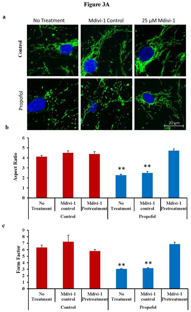
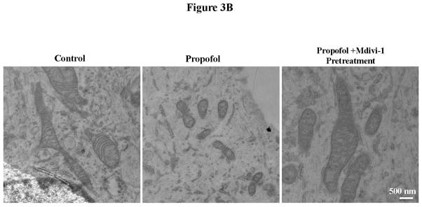
Propofol exposure induced severe mitochondrial fragmentation in human embryonic stem cell (hESC)-derived neurons. (A) hESC-derived neurons were exposed to 6 hours of 20 μg/mL propofol or the vehicle control and immunostaining with the TOM20 (translocase of outer mitochondrial membranes 20 kDa) antibody was used to visualize the mitochondria. The cells were then imaged on the confocal microscope to assess the mitochondrial morphology. The mitochondria appeared severely fragmented following propofol exposure and this was rescued by pretreatment of the cells for 1 hour with 25 μM mdivi-1 (a). The images were analyzed using ImageJ to assess the form factor (mitochondrial branching) and the aspect ratio (mitochondrial length). The aspect ratio was significantly reduced in the propofol-treated group indicating mitochondrial fragmentation. This was rescued by pretreatment of the cells for 1 hour with 25 μM mdivi-1 (b). The form factor was also significantly reduced in the cells treated with propofol alone or propofol with mdivi-1 control pretreatment when compared to all control-treated groups, further indicating increased mitochondrial fission. The form factor was rescued to control levels in cells pretreated for 1 hour with 25 μM mdivi-1 prior to propofol exposure (c). (B) hESC-derived neurons exposed to propofol or control conditions with or without mdivi-1 pretreatment were fixed and imaged using electron microscopy to visualize the cellular ultrastructure and mitochondrial morphology. The mitochondria of the control treated group were large and elongated with clear cristae and appeared generally healthy. In contrast, the mitochondria in the propofol-treated group were extremely small with disorganized and abnormal inner mitochondrial membranes. These mitochondrial abnormalities were not present in the propofol-treated group when the cells were pretreated with the mitochondrial fission blocker, mdivi-1. (**P < 0.01 vs. all control groups and propofol mdivi-1 pretreatment group, n = 5 coverslips/group).


