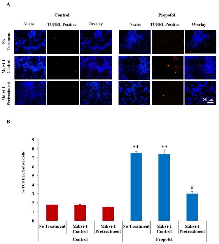
Fig. 4.
Propofol induced cell death was attenuated by pretreatment of the cells with the mitochondrial fission blocker, mdivi-1. (A) TUNEL staining was used to assess cell death in human embryonic stem cell (hESC)-derived neurons. Cells were exposed for 6 hours to 20 μg/mL propofol or the vehicle control following a 1 hour pretreatment with media alone (no treatment), the mdivi-1 vehicle control (mdivi-1 control) or 25 μM mdivi-1 (mdivi-1 pretreatment) and stained using the TUNEL kit. Most of the TUNEL staining (red dots) co-localized with the nuclei stain (blue dots stained with Hoechst 33342) and there were more TUNEL positive cells observed in the “propofol no treatment” and “propofol midivi-1 control” treated groups when compared to all other groups. (B) The number of TUNEL-positive cells and total cell nuclei present in the field were manually counted to obtain a percent of TUNEL-positive cells. The percent of TUNEL-positive cells was significantly increased in the “propofol no treatment” and “propofol mdivi-1 control” treated groups when compared to all control-treated groups indicating increased cell death. The increase in cell death was attenuated by mdivi-1 pretreatment. (**P < 0.01 vs. all control groups and propofol mdivi-1 pretreatment group, # P < 0.01 vs. dimethyl sulfoxide (DMSO). mdivi-1 pretreatment group, n = 5 coverslips/group). TUNEL = terminal deoxynucleotidyl transferase-mediated deoxyuridine triphosphate in situ nick end labeling.

