
Figure 4.
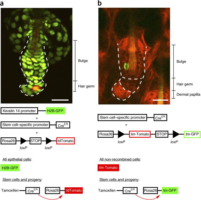
Labeling hair follicle stem cells with inducible Cre reporters. These images demonstrate the diversity of reporters that can be used to target specific cell populations, including stem cells, within the hair follicle. (a) K14H2BGFP;tdTomato;Lgr5CreER hair follicle and, directly below, a corresponding schematic describing the dual fluorescent reporter/Cre recombinase system. Upon Cre recombination, the fluorescent tag switches from nuclear green to cytoplasmic tomato (red). The cycling portion of the follicle is outlined (dotted line) and divided into bulge and hair germ, and all epithelial nuclei are in green. A single tdTomato Cre-recombined epithelial cell is in red. (b) mTmG;K19CreER hair follicle and, directly below, a corresponding schematic explaining the mTmG/Cre recombinase system. The cycling portion of the follicle is outlined (dotted line) and divided into bulge, hair germ and dermal papilla. All epithelial nuclei contain a red membrane label, and the single Cre-recombined cell contains a green membrane label. Both images demonstrate single-cell labeling as a result of low induction of the Cre recombinase system through administration of a low dose of tamoxifen. Scale bars, 20 μm. All studies and procedures involving animal subjects were approved by the Institutional Animal Care and Use Committee at the Yale University School of Medicine and conducted in accordance with the approved animal handling protocol.