
Figure 5.
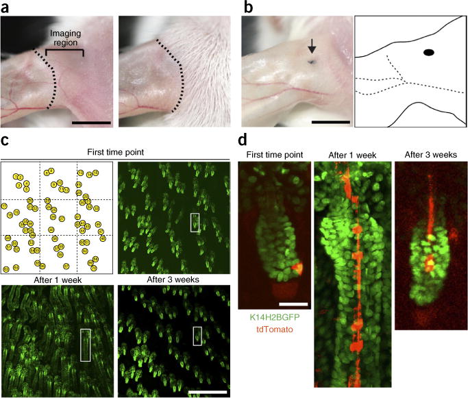
Strategy for repeated imaging of the same skin region. (a) Magnified views of the dorsal ear skin immediately after depilation (left) and 10 d later (right). The dotted line indicates the borders between areas of active hair growth and quiescence. Imaging should be done in the ear region to the right of the dotted line, indicated by a bracket, where hair is actively growing. Scale bar, 5 mm. (b) A punctate tattoo (indicated by the arrow) can be used for creating additional landmarks, which, together with the distinctive pattern of the vasculature network, can be used for navigating back to the same region for subsequent imaging sessions. Scale bar, 5 mm. (c) Digital tiling can be used to identify and revisit the same hair follicles in subsequent imaging sessions. Sequential views of the same skin region, showing hair follicles in different stages of the hair cycle (telogen I, upper right; anagen II, lower left; telogen II, lower right). Scale bar, 0.5 mm. The white box highlights a single follicle magnified in d. Snapshots of a single K14H2BGFP;tdTomato follicle at three different time points during the regeneration cycle, in which a single stem cell is labeled and traced. All studies and procedures involving animal subjects were approved by the Institutional Animal Care and Use Committee at the Yale University School of Medicine and conducted in accordance with the approved animal handling protocol.