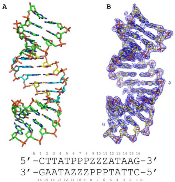
Figure 2.
Crystal structure of 16-mer oligonucleotide containing six consecutive Z:P pairs (3/6 ZP) is shown in (A) as a stick rendering with C, green for non-Z:P pairs, yellow for Z, and cyan for P, O in red, nitrogen in blue, phosphorous in orange. (B) The experimental electron density map derived from Br SAD phasing is shown as a blue mesh contoured at 1.5 σ with the final refined structural model as a stick model. The sequence of the oligonucleotide is shown along with numbering scheme employed in the coordinate file.

