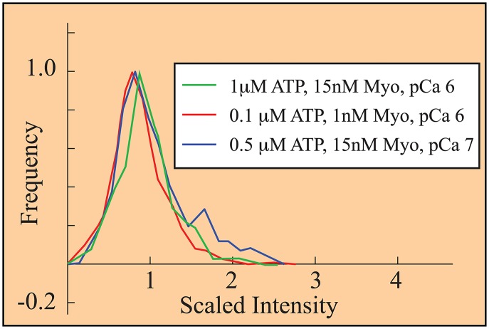
Fig 6. Histograms, collected under three different experimental conditions [52], collapse upon rescaling fluorescence.
Scaled intensity, defined in the text, gives a single fluorescently-tagged myosin (GFP-S1) an intensity of 1. The observation that each histogram has a peak near 1 suggests that 1) under these three conditions, mostly single GFP-S1s bind; and 2) the emission of an excited GFP is constant and measured fluorescence is inversely proportional to frame rate. This latter is a central assumption of our analysis.

