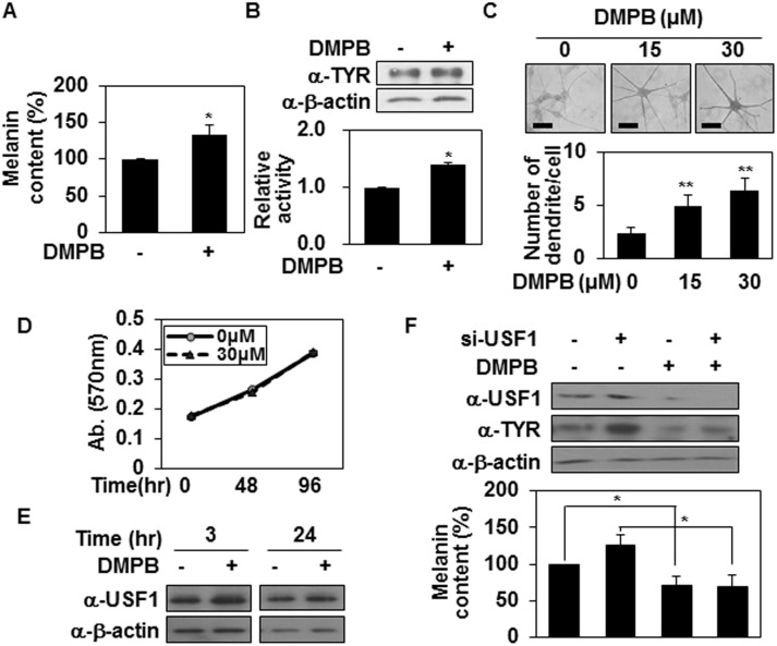
Fig 5. DMPB promotes melanin synthesis in human melanocytes.
(A, B) Human melanocytes were treated with 30 μM of DMPB. After 48 hr melanin contents were analyzed by measuring the absorbance at 405 nm (A). The mean percentages of melanin content are shown *, p < 0.05 versus DMSO treated cells. Tyrosinase expression levels were analyzed by Western blot analysis (B; top panel). Cell lysates (100 μg) were reacted with L-DOPA at 37°C for 2 hr, and tyrosinase activity was determined at 470 nm (B; bottom panel). The mean percentages of tyrosinase activity ± SD are shown. *, p < 0.05 versus DMSO treated cells. (C) Human melanocytes were plated on 12-well plates, treated with the indicated concentrations of DMPB for 48 hr, and reacted with L-DOPA at 37°C for 30 min. Bright-field microscopic images are shown (top panel). Scale bars = 50 μm. The mean percentages of the number of dendrite/cell ± SD are shown (bottom panel). **, p < 0.01 versus DMSO treated cells. (D) Primary melanocytes were incubated with DMPB (30 μM) in a 96-well plate for the indicated periods, and cell viability was determined by MTT-based spectrophotometric assay. Percentage values were compared between treated and untreated (control) cells. The data are expressed as mean ± SD from three independent experiments. (E) Primary melanocytes were treated with 30 μM of DMPB for the indicated periods. USF1 protein levels were analyzed by Western blotting. (F) Primary melanocytes were transfected with USF1 targeting siRNA and treated with DMPB (30 μM). After 48 hr, tyrosinase and USF1 expression levels were analyzed by Western blot analysis (top panel). Cells were harvested and melanin contents were analyzed by measuring the absorbance at 405 nm (bottom panel). The mean percentages or melanin content are shown *, p < 0.05.

