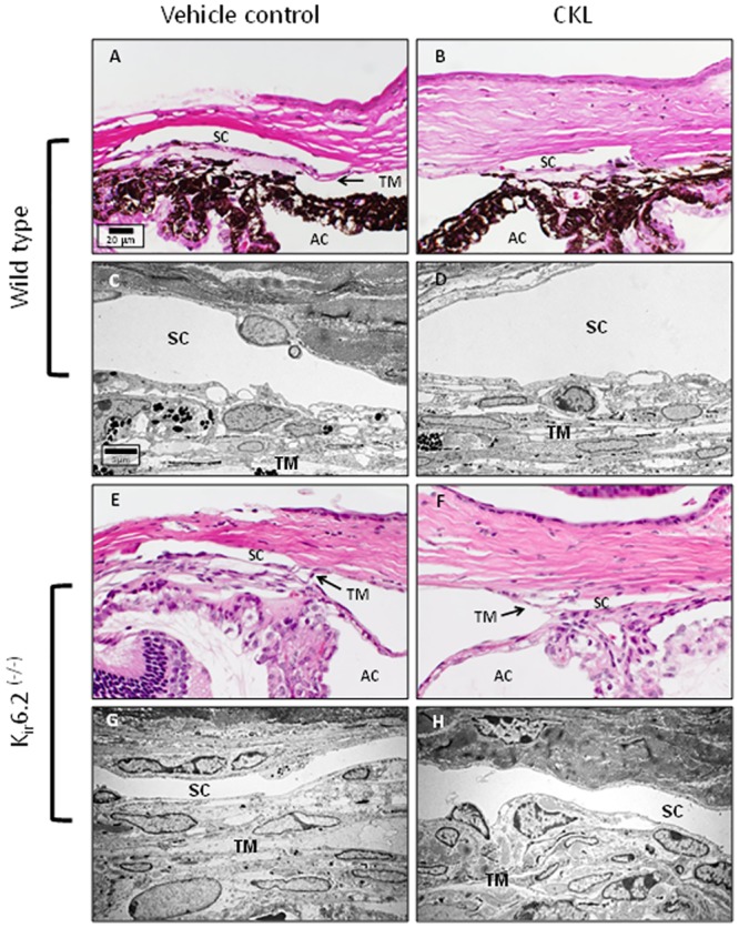
Fig 4. Representative images of the conventional outflow pathway of C57BL/6 (A-D) and Kir6.2(-/-) (E-H) treated and vehicle control mice.

Similar to human anterior segments, comparison between vehicle and cromakalim treated eyes showed no observable cell or tissue changes. Both vehicle and cromakalim treated groups showed normal morphology and ultrastructure with intact inner and outer walls of Schlemm’s canal, viable cells and an evenly distributed extracellular matrix. SC = Schlemm’s canal; TM = trabecular meshwork; AC = anterior chamber.
