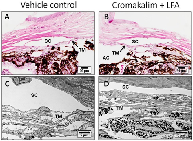
Fig 6. Representative transmission electron micrographs of mouse eyes treated with cromakalim + latanoprost free acid (LFA) or vehicle.

Comparison of the micrographs show normal anatomy and ultrastructure in vehicle (A, C) and cromakalim + LFA treated tissue (B, D), indicating no observable changes due to treatment. SC = Schlemm’s canal; TM = trabecular meshwork; AC = anterior chamber.
