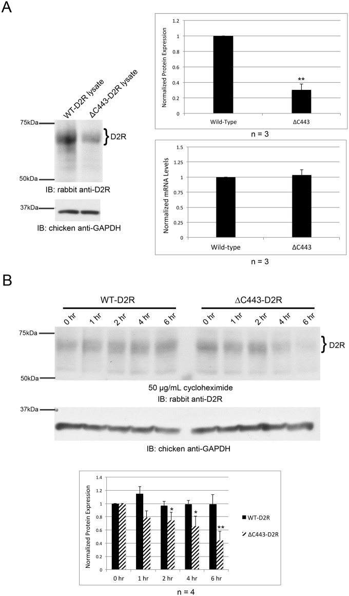
Fig 7. Deletion of C443 affects stability of the D2R.
(A, B) FLAG-tagged WT-D2R or ΔC443-D2R cDNAs were transiently expressed in HEK-293T cells. Quantitation of receptor expression was normalized to GAPDH expression for protein and mRNA. (A) Proteins were separated by SDS-PAGE and analyzed by Western blotting (left). WT-D2R and ΔC443-D2R mRNA expression levels were determined by RT-qPCR and normalized to wild-type levels (bottom graph). The top bar graph represents the average pixel density (as determined by ImageJ) from three separate experiments. All data were analyzed using a two-sided unpaired Student’s t test (expressed as ± SEM, n = 3, **P<0.01). (B) Cells were treated with 50 μg/mL cycloheximide for the indicated times. Proteins were separated by SDS-PAGE and analyzed by Western blotting (top). The bar graph represents the average pixel density (as determined by ImageJ) from four separate experiments. Data were analyzed using a two-sided unpaired Student’s t test (expressed as ± SEM, n = 4, *P < 0.05, **P<0.01).

