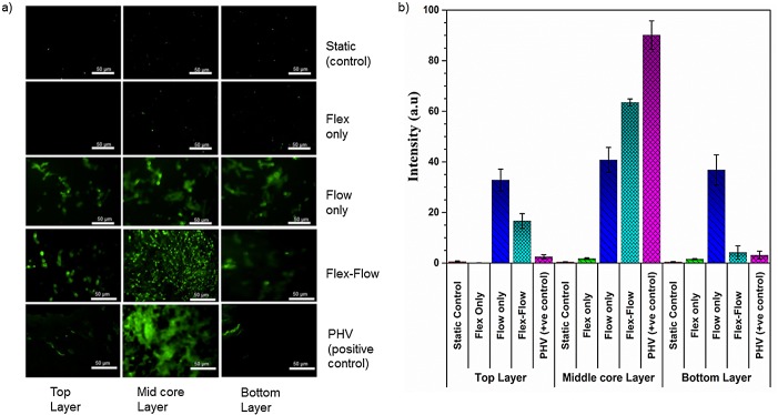
Fig 4.
a) Immunofluorescence staining of α-SMA protein on both surface layers (~90μm thickness on each side), middle core (interstitial tissue) regions (~400 μm thickness) of the valve; 1st row: Static Controls; 2nd row: Flex; 3rd row: Flow; 4th row: Flex-Flow conditioning; 5th row: porcine heart valve as Positive control. Among the experimental groups, α-SMA-expressing cells were found to be predominant within the interstitial region (middle layer) of the engineered tissues in solely the Flex-Flow group; b) Quantification of positive staining (green; from images in part a) for α-SMA signal-intensity in four experimental groups; Samples exposed to flex-flow expressed a significantly higher level of positive α-SMA (p < 0.05) in comparison to the control group. PHV was treated as the positive control.

