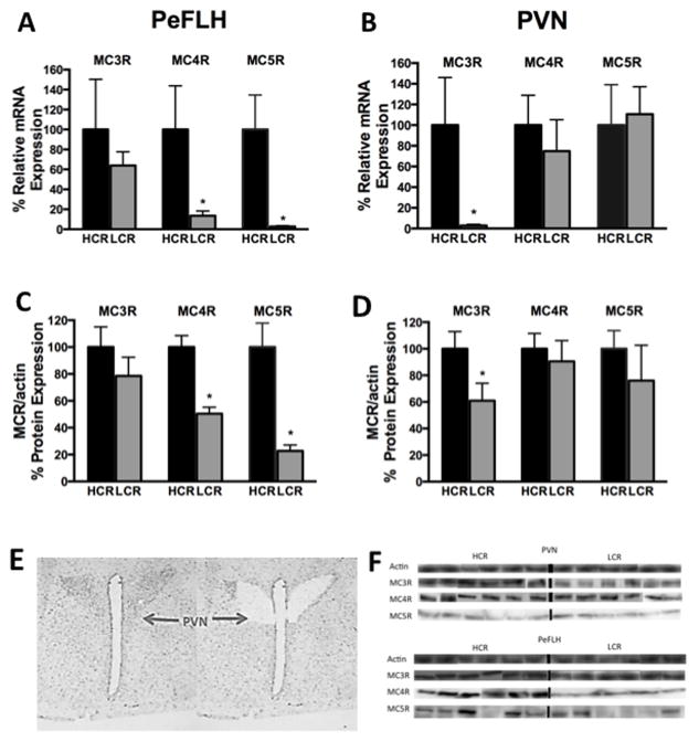
Figure 1. Melanocortin receptor expression in hypothalamic nuclei.
Region-specific elevations in melanocortin receptors subtypes in the perifornical lateral hypothalamus (PeFLH) and the paraventricular nucleus (PVN) in lean, active high-capacity rats (HCR) compared to low-capacity rats (LCR) measured by quantitative PCR (Q-PCR) and Western blots. (A) Heightened levels of melanocortin receptors 4 and 5 (MC4R, MC5R) were seen in HCR in PeFLH. (B) In the PVN, melanocortin 3 receptor (MC3R) was higher in HCR. This pattern was also seen in protein expression (C) and (D). (E) Photomicrographs of the hypothalamus before and after laser capture microdissection of the PVN. (F) Representative blots of MC3R, MC4R, and MC5R from micropunched samples of HCR and LCR from PVN and PeFLH with actin as the loading control. *p<0.05, different than HCR for the same receptor and brain region. N= 8–12 per group, data represent mean plus SEM. Immunoblots for each protein subtype represent bands from the same experiment and may have been spliced to reorder and show parallel comparisons between MC3, 4, and 5 receptors within the same animal.

