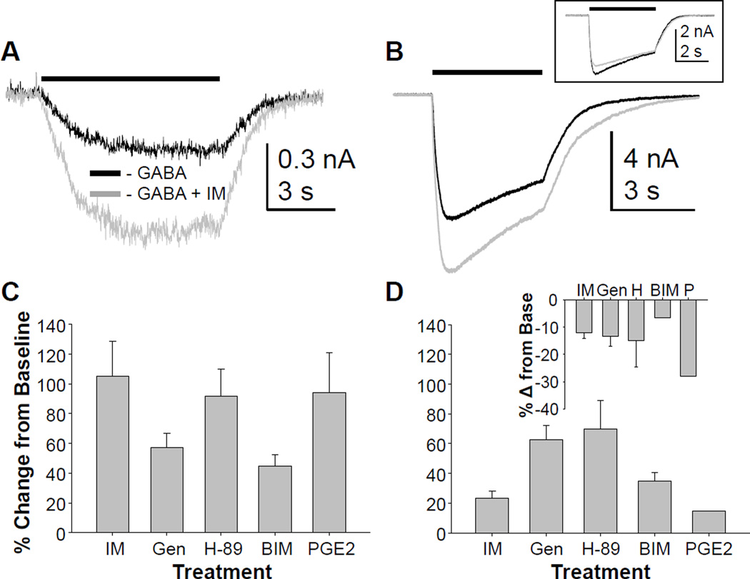
Figure 5.
High and low affinity GABAA currents in human sensory neurons are modulated by inflammatory mediators. A. GABA was applied before and then 3 minutes after the application of a combination of inflammatory mediators (IM). In the majority of neurons tested (15 of 16), the high affinity current (evoked with 10 µM) GABA was increased following IM application. B. Changes in low affinity currents were studied in the same neurons, and in half of these (6 of 13), the low affinity current was also increased by IM. Inset: In a minority of neurons (3 of 13), however, a decrease in low-affinity current was observed. Separate groups of neurons were pretreated with the TK kinase inhibitor, genistein (Gen, n = 11), the PKA inhibitor H-89 (n = 8), or the PKC inhibitor BIM (n = 5) prior to the application of GABA and then GABA with inflammatory mediators. Pooled high (C) and low (D) affinity current data were analyzed as a percent change from the baseline GABA current evoked from neurons treated with IM alone or pre-treated with kinase inhibitors. A two-way ANOVA indicated a significant interaction between current type (high vs low affinity) and treatment. Of note, only the neurons in which low affinity current increased by > 10% were included in this analysis, and plotted in panel D. The IM-induced decrease in low affinity current (D inset) was observed in a subpopulation of neurons pre-incubated with each inhibitor and no significant impact of inhibitor was detected in these neurons. Finally, a group of neurons (n = 5) was treated with PGE2 (P) alone, rather than in combination with bradykinin and histamine. Pooled high (C) and low (D) affinity current data were included with plots of pooled IM data for comparison.

