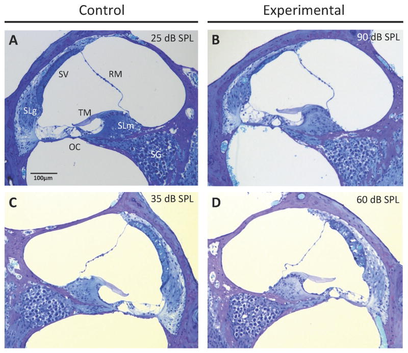
Figure 2.
Cochlear duct histomorphology. Light microscopic images of positive control (A, C) and experimental (B, D) mice at 12 months of age. RM, Reissner’s membrane; SV, stria vascularis; SLm, spiral limbus; TM, tectorial membrane; SLg, spiral ligament; OC, organ of Corti; SG, spiral ganglion. Click ABR threshold values for the corresponding ear are shown for each section. (B) Atrophy of the stria vascularis was observed in experimental ears with severe hearing loss. (D) The stria vascularis was edematous in experimental ears with less severe hearing loss.

