Figure 6.
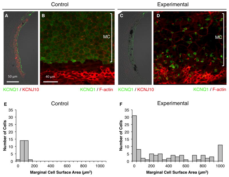
Stria vascularis protein expression. Representative immunostaining of KCNQ1 (green) and KCNJ10 (red) in cryosections (A, C) or KCNQ1 (green) and F-actin (red) in whole-mounted specimens (B, D) of control (A, B) or experimental stria vascularis (C, D) in the middle turn. n = 6 ears for control mice, and n = 12 ears for experimental mice. MC, marginal cells. KCNQ1 fluorescence was reduced and KCNJ10 staining was absent in experimental ears at 12 months of age. Some marginal cells were expanded and lacked detectable KCNQ1, while others were abnormally small with KCNQ1 concentrated or aggregated within small cell surfaces (D). Apical surface areas of marginal cells for 3 control ears (E; n = 30 cells) and 3 experimental ears (F; n = 96 cells). Bin area = 50 μm2. If the surface area was > 1050 μm2, it was included in the 1000–1050 μm2 bin.

