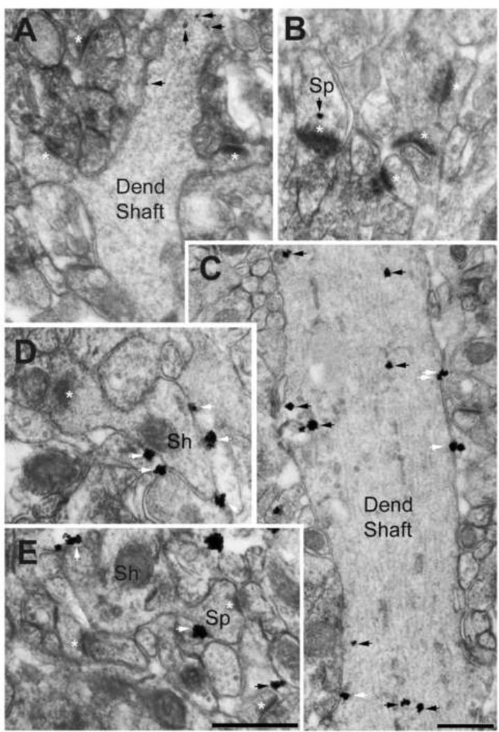
Figure 2. Electron micrographs showing α4-immunoreactivity in dendritic shafts and spines of the dorsal hippocampal CA1 of adolescent female mice that received P4 injection.
Panels A and B were taken from an animal categorized as vulnerable to ABA, while panels C, D and E were taken from an animal categorized as resistant to ABA. Animals had undergone two ABA inductions and were of age PND59. Black arrows point to cytoplasmic location of silver-intensified gold (SIG) particles reflecting α4-immunoreactivity, while white arrows point to SIG particles located at the extracellular surface of plasma membranes. Note the paucity of SIG particles in the dendritic shaft cytoplasm of the vulnerable animal (Panel A), compared to the dendritic shaft of the resistant animal (Panel C). Moreover, plasma membrane location of SIG particles is readily apparent in the dendritic shaft of the resistant animal (Panels C and ‘Sh’ in D), but not of the vulnerable animal. Similarly, SIG is sparse in spines of the vulnerable animal (Sp, white asterisks point to postsynaptic densities, Panel B) but more numerous and located more often at the plasma membrane of the resistant animal (panel E). The black arrow at the lower right corner of panel E points to cytoplasmic labeling in an astrocytic process immediately adjacent to an axo-spinous synapse with a PSD. Calibration bar in E = 500 nm and also applies to panels A, B, and D, all of which were captured at a magnification of 40,000x using AMT Camera system and Hamamatsu's CCD camera. Calibration bar in C = 500 nm, captured at a magnification of 40,000x.

