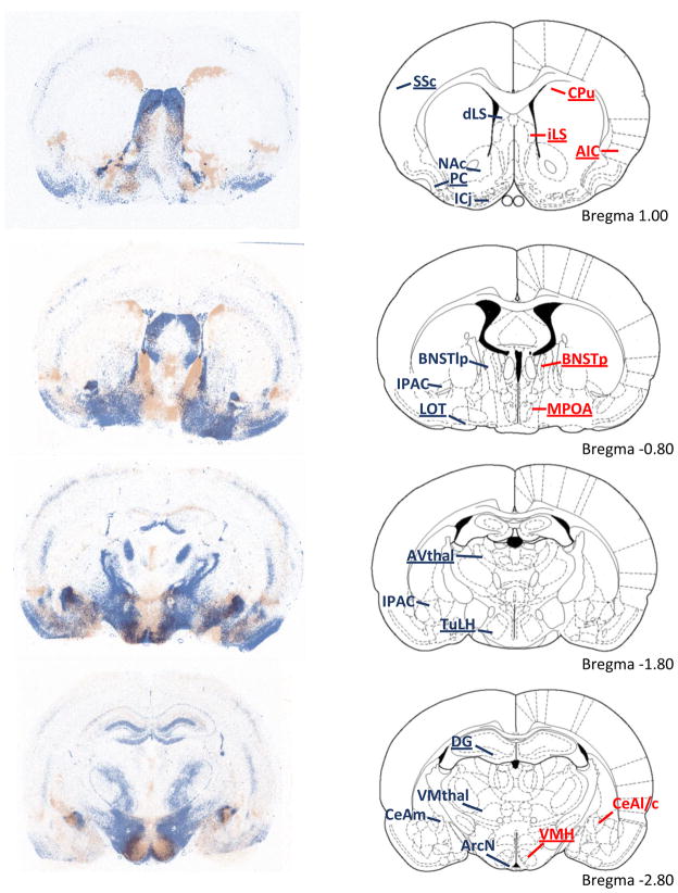
Fig. 3.
Overlay of V1aR binding densities (blue) and OTR binding densities (red) in forebrain areas of male Wistar rats from adjacent coronal sections. The right column depicts representative rat brain images adapted from The Rat Brain Atlas (Paxinos & Watson, 1998). Overall, there is little overlap between V1aR binding and OTR binding profiles. Dense V1aR binding is found in the somatosensory cortex (SSc), piriform cortex (PC), Islands of Calleja (ICj), nucleus accumbens (NAc), lateral septum (LS), lateral dorsal BNST (not shown), lateral posterior BNST (BNSTlp), medial posterior BNST (not shown), nucleus of the lateral olfactory tract (LOT), dentate gyrus (DG), tuberal lateral hypothalamus (TuLH), anteroventral thalamus (AVthal), suprachiasmatic nucleus of the hypothalamus (not shown), interstitial nucleus of the posterior limb of the anterior commissure (IPAC), arcuate nucleus of the hypothalamus (ArcN), ventromedial thalamus (VMthal), and medial central amygdala (CeAm). Dense staining of OTR binding is found in the dorsal caudate putamen (CPu), agranular insular cortex (AIP), posterior BNST (BNSTp), medial preoptic area (MPOA), ventral medial hypothalamus (VMH), and lateral and capsular central amygdala (CeAl/c). Regions which show sex differences in V1aR or OTR binding are underlined.

