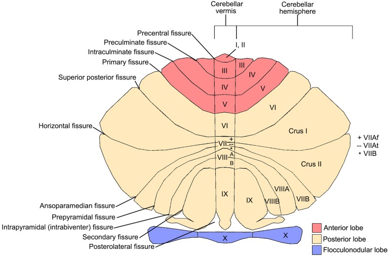
Figure 1.
Cerebellar anatomy showing major fissures, lobes, and lobules. The cerebellum is flattened to show the anterior lobe (red; lobules I-V), posterior lobe (cream; lobules VI-IX), and flocculonodular lobe (purple; lobule X). The 10 cerebellar lobules (labeled I-X) are labeled in both the vermis and hemispheres. Lobule VII is subdivided into Crus I, Crus II, and VIIB in the hemispheres, and VIIAf, VIIAt, and VIIB in the vermis. Lobule VIII is subdivided into VIIIA and VIIIB. Figure courtesy of Professor Jeremy Schmahmann, Massachusetts General Hospital, Boston.

