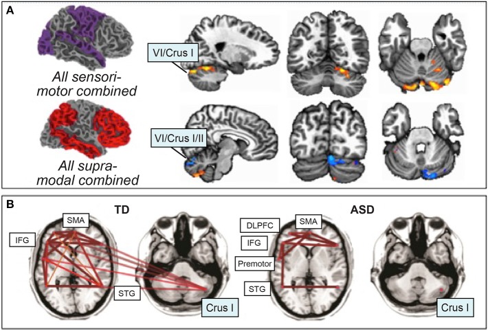
Figure 3.
Resting-state functional connectivity in ASD. (A) Atypical increased functional connectivity between sensorimotor regions of the cerebral cortex and cerebellar lobules VI and VII (orange), decreased functional connectivity between supra-modal association cortices and lobules VI and VII (blue). Orange, ASD greater rsFC than typically-developing; blue, ASD less rsFC than typically-developing. Figure adapted with permission from Khan et al. (2015). (B) Preserved functional connectivity in ASD between supratentorial language regions, in contrast with the lack of cerebro-cerebellar connectivity between right Crus I/II and left-hemisphere language regions. Figure adapted from Verly et al. (2014).

