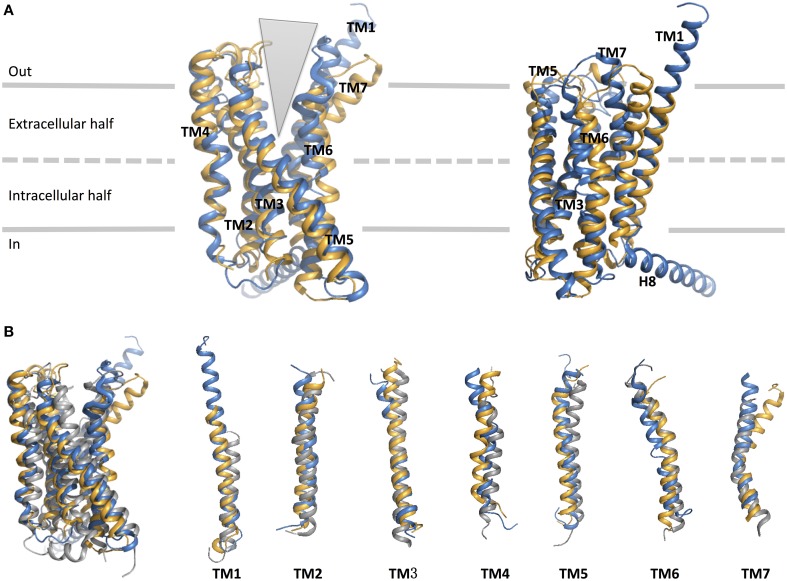
Figure 9.
Structural alignment of CRF1 (gold, PDB ID 4K5Y) and GPCR (blue, PDB ID 4L6R). (A) The receptors are viewed from two different angles from within the membrane. The TM helices are labeled and comprise the two halves of the V-shape open configuration. (B) Structural comparison of the two family B GPCRs with a family A GPCR, dopamine D3 receptor (gray, PDB ID 3PBL), in its inactive form. Individual TM helices are shown after superposition of the three receptors.

