
FIGURE 2.
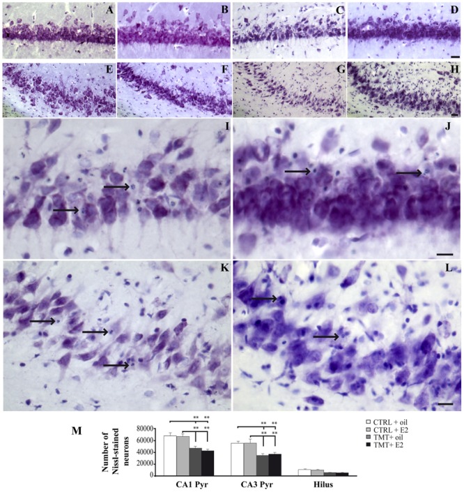
Trimethyltin-induced hippocampal damage. (A–L) Representative micrographs of Nissl-stained rat hippocampal sagittal sections from CA1 (A–D,I,J) and CA3 subfields (E–H,K,L) of CTRL + oil- (A,E), CTRL + E2- (B,F), TMT + oil- (C,G,I,K), TMT + E2- (D,H,J,L) treated rats. Neuronal loss and apoptotic bodies are clearly detectable in CA3 and CA1 pyramidal neurons (arrows in I–L) of both TMT + oil- (C,G,I,K) and TMT + E2- (D,H,J,L) treated animals. Scale bar: 80 μm in (A–H), 40 μm in (I–L). (M) Bar graphs indicate quantitative analysis of Nissl-stained neurons in CA1 and CA3 pyramidal cell layer and hilus of the different experimental groups. A significant reduction in the number of Nissl-stained cells is evident in CA1 and CA3 pyramidal layers of both TMT-treated-groups compared with control groups. No differences are detectable between the two groups of TMT-treated rats. The values are given as means ± SE (∗∗p < 0.001).