FIGURE 4.
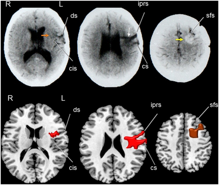
(Top) Axial CT of patient RC showing a hypodense lesion (middle image) involving the middle part of the left precentral gyrus (white arrow) and extending deeply to dorsal head of the caudate nucleus. Note in the left image the involvement of the anterior insula (red arrow) and shrinkage of the middle and posterior insular cortex (orange arrow) and part of the inferior precentral gyrus and superior temporal gyrus. The right image shows a hyperdense rounded image which corresponds to a remnant of the arterio-venous malformation (AVM) involved the left superior frontal gyrus reaching the pre-supplementary motor area and the subcortical white matter (anterior centrum semiovale; yellow arrow). (Bottom) Drawings were made using MRIcro software (Rorden, C., 2005. www.mccauslandcenter.sc.edu/mricro/mricro/) on T1-weighted axial images. The lesion is drawn in red (left and middle images) and the AVM in brown (right image). Fissures and sulcus are indicated with white arrows. ds indicates: diagonal sulcus; cis: central insular sulcus; iprs: inferior precentral sulcus inferior; cs: central sulcus (sulcus of Rolando); and sfs: superior frontal sulcus. Terminology and abbreviations for fissures and sulci were taken from the atlas of neuroanatomy of language regions of the human brain (Petrides, 2014). R: indicates right and L: left.

