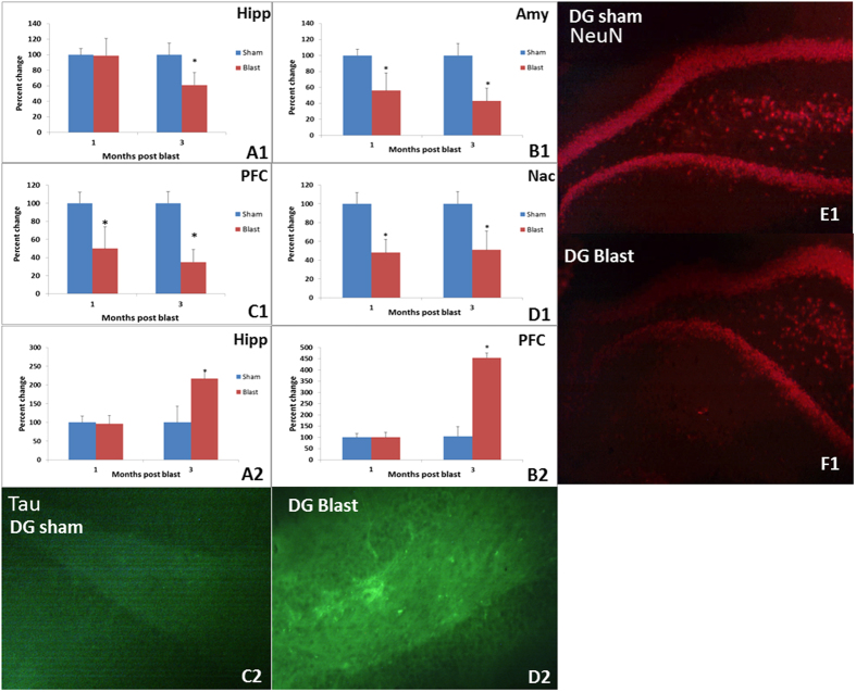
Figure 4.
Top panels: Staining for NeuN, a protein found exclusively in mature neurons, was decreased in Amy (B1), Hipp (A1), Nac (D1), and PFC (C1) of blast animals (*p < 0.05). Representative immunochemical staining for NeuN (red) in the DG of control (E1) and blast groups (F1) are shown. Bottom panels: Increased tau pathology was observed in Hipp (A2) and PFC (B2) obtained three months after blast, when compared to control group (*p < 0.05). Representative immunochemical staining for tau (green) in DG from control (C2) and blast group animals at 3 months (D2) are shown.

