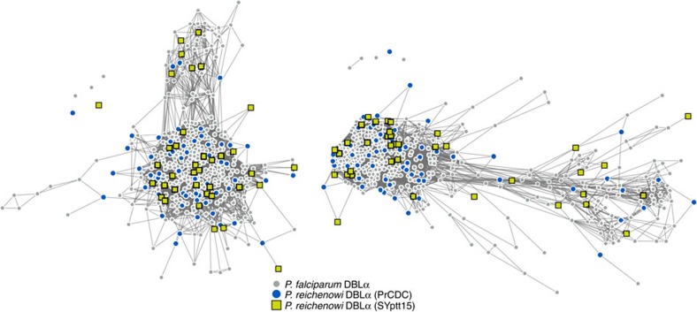
Figure 2. Networks of DBLα sequences from P. reichenowi and P. falciparum.
Each node represents a DBLα HVR sequence and each link represents a shared amino-acid substring of significant length21. Laverania species and strain origin is indicated by node colour and shape. Left and right networks correspond to left and right HVRs, respectively. P. falciparum and P. reichenowi sequences do not cluster by species or sample in either HVR. Link lengths and node placements are determined by a force-directed layout to better reveal structure, if it exists (see the Methods section). Additional analyses of these networks are shown in Supplementary Fig. 1.

