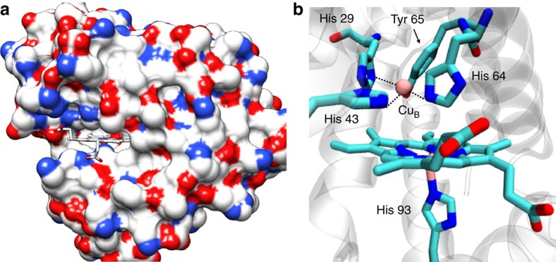
Figure 1. Crystal structure.
Crystal structure of a Mb-based biosynthetic model of CcO, F33Y-CuBMb; pdb id: 4FWY. (a) The haem cofactor is in a cleft on the molecule protein surface, (colour coded according to the charge of the residues), with the propionate groups exposed to the solvent. (b) The computer model of G65YCuBMb showing its catalytic centre containing the distal CuB bound to histidines and a tyrosine 65.

