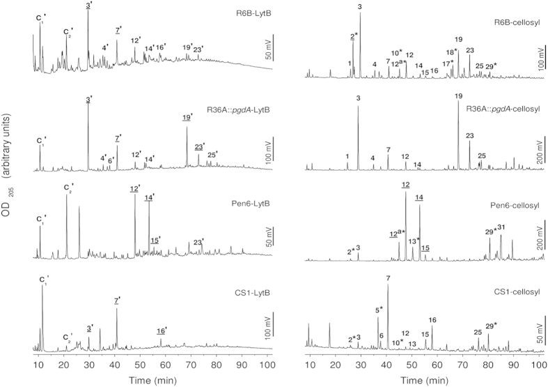
Figure 3. Comparison of the pattern of PG fragments released with LytB or cellosyl from cell wall or PG from different pneumococcal strains.
Reduced PG fragments solubilized by PG-digestion with cellosyl or cell-wall digestion with LytB were separated on a Prontosil C18 column, and the OD205 of the eluate was monitored. Peaks obtained with cellosyl, used as a control of PG muropeptide composition, are numbered as in Bui et al. 20127. Corresponding structures of the LytB-released fragments (marked with apostrophes) have similar retention time but differ in the sugar at the reducing end (N-acetylglucosaminitol for LytB vs. N-acetylmuramitol for cellosyl; see Supplementary Fig. S4). PG fragment structures are shown in Supplementary Fig. S3. Labels with asterisks indicate deacetylated muropeptides and those underlined correspond to peaks analyzed by MS (see Supplementary Table S2). The structures corresponding to C1-C2 peaks could not be determined due to the co-elution of phosphate from the sample buffer, which interferes with ESI-MS/MS analysis.

