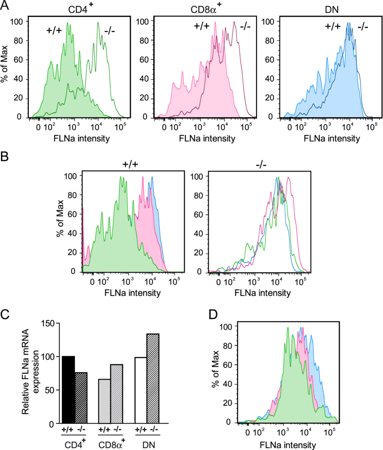
Figure 3. ASB2α mediates FLNa degradation in cDC subsets.
(A,B) Expression of FLNa was assessed by intracellular flow cytometry coupled to extracellular flow cytometry in CD11chiCD4+CD8α− (CD4+; green), CD11chiCD4−CD8α+ (CD8α+; pink) and CD11chiCD4−CD8α− (DN; blue) spleen cDC subsets from Mx1-Cre (+/+) and Mx1-Cre;ASB2fl/fl (−/−) mice that have received poly(I·C). Filled histograms show expression of FLNa in cDC subsets of control mice whereas unfilled histograms show expression of FLNa in cDC subsets of Mx1-Cre;ASB2fl/fl mice that have received poly(I·C) (sample size: +/+ = 3; −/− = 4). One representative experiment out of three is presented. (C) Relative expression of FLNa mRNA assessed by RT-qPCR in CD4+, CD8α+ and DN cDC subsets in spleen of Mx1-Cre (+ /+ ) and Mx1-Cre;ASB2fl/fl (− /− ) mice that have received poly(I·C) (sample size: +/+ = 6; −/− = 6). (D) Expression of FLNa was assessed by intracellular flow cytometry coupled to extracellular flow cytometry in CD4+, CD8α+ and DN cDCs isolated from mesenteric lymph nodes of ASB2+/+ mice (sample size = 5). One representative experiment out of three is presented.

