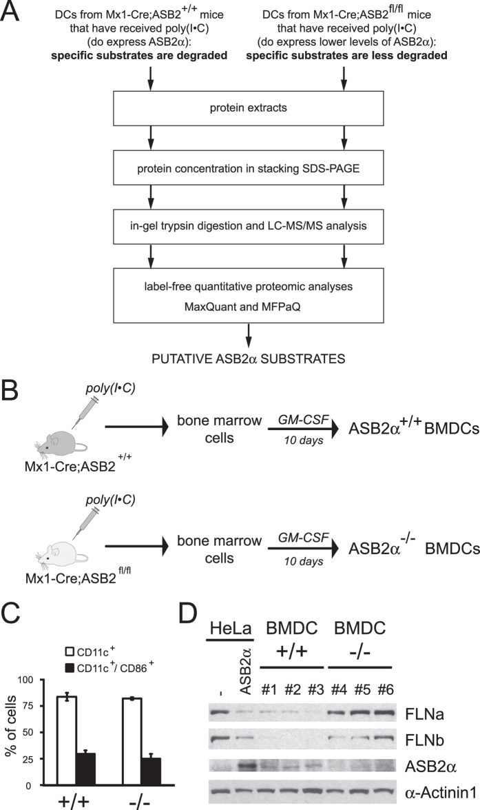
Figure 5. Experimental design for identification of ASB2α substrates in DCs.
(A) Experimental design. (B) Experimental outline of ASB2 deletion and generation of GM-CSF BMDCs. (C) Expression of CD11c and CD86 at the cell surface of ASB2α−/− and ASB2α+/+ BMDCs. (D) Expression of FLNa, FLNb, ASB2α, α-actinin 1 was analyzed by western blot using 10-μg aliquots of protein extracts of ASB2α−/− and ASB2α+/+ BMDCs from three independent experiments and 3-μg aliquots of cell extracts of HeLa cells transfected with a mouse ASB2α expression vector (ASB2α) or mock-transfected (−). The drawing in panel B is from P.G.L.

