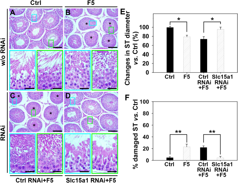
Figure 6. Effect of Slc15a1 knockdown on F5-peptide induced impairment of spermatogenesis and germ cell loss in rat testes.
(A) Representative photographs of paraffin sections of testes stained with hematoxylin and eosin. Results of HE staining were shown in normal testis (Ctrl, A), testis treated with F5-peptide at 320 μg/testis (F5, B), or testis pre-transfected with non-targeting control siRNA (Ctrl RNAi+F5, C) or Slc15a1 siRNA (Slc15a1 RNAi+F5, D) of a group of adult rats (~300–350 gm b.w.) with n = 3 rats in each treatment group. Scale bar in A–D = 150 μm. The “blue” and “green” encircled micrographs are magnified images of the corresponding boxed areas of the lower magnification, bar = 50 μm. (E,F) Morphometric changes of seminiferous tubules (ST) (n = 200 tubules from 3 rat testes). The shrinkage of tubules indicated by changes in ST diameter (the average ST diameter in treatment group vs. that in its corresponding control) (E), and the percentage of damaged ST number manifested by germ cell exfoliation (annotated by asterisks in tubules seen in B,C) in treatment group vs. that in its corresponding control (F) are summarized in the bar graphs (E,F), respectively. *P < 0.05, **P < 0.01 when compared to normal control rats.

