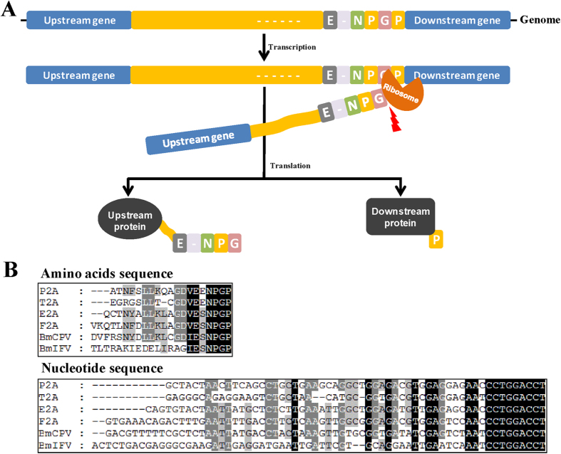
Figure 1. Cleavage mechanism and sequence analysis of 2A self-cleaving peptide.
(A) Self-cleavage mechanism of 2A self-cleaving peptide. The cleavage site locates between glycine (G) and praline (P) at its C-terminus. After self-cleavage, amino acids residues at the N-terminus of 2A linked to upstream protein and its last praline residue remained to the downstream protein, but the residues of 2A could be removed through furin and signal peptide9. (B) Sequence analysis of six 2As which was respectively from porcine teschovirus-1 2A (P2A), thoseaasigna virus 2A (T2A), equine rhinitis a virus 2A (E2A), foot and mouth disease virus 2A (F2A), cytoplasmic polyhedrosis virus 2A (BmCPV2A) and flacherie Virus 2A (BmIFV2A).

