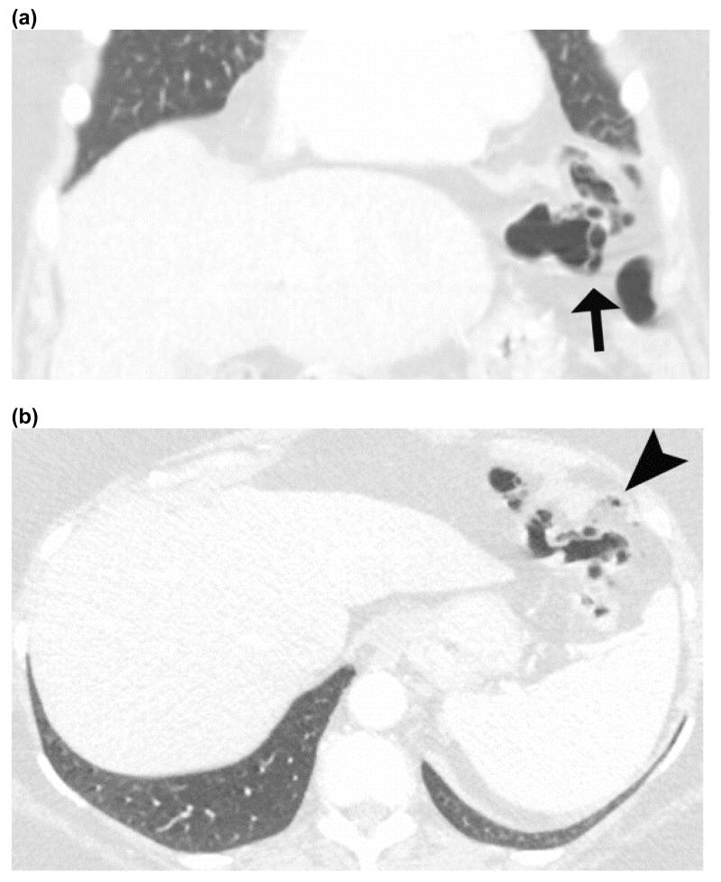
Figure 10.
Benign pneumatosis – CT images in lung window of a patient who presented with abdominal pain after trauma. Coronal image (a) shows pneumatosis cystoides coli (black arrow), while the axial images (b) shows small volume of pneumoperitoneum (black arrow head). Patient was admitted for observation and discharged without any intervention as his pain resolved spontaneously.

