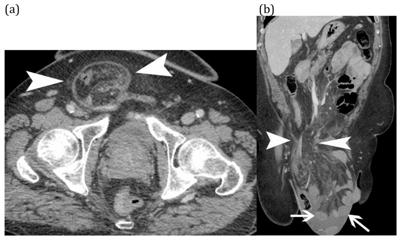
Figure 13.
Strangulated small bowel – Axial (a) and coronal (b) CT images of incarcerated small bowel (white arrows) in a large right inguinal hernia (white arrow heads). CT findings include hypoenhancement of the small bowel wall (arrows) with adjacent fluid and fat stranding of the associated mesentery in the hernia sac.

