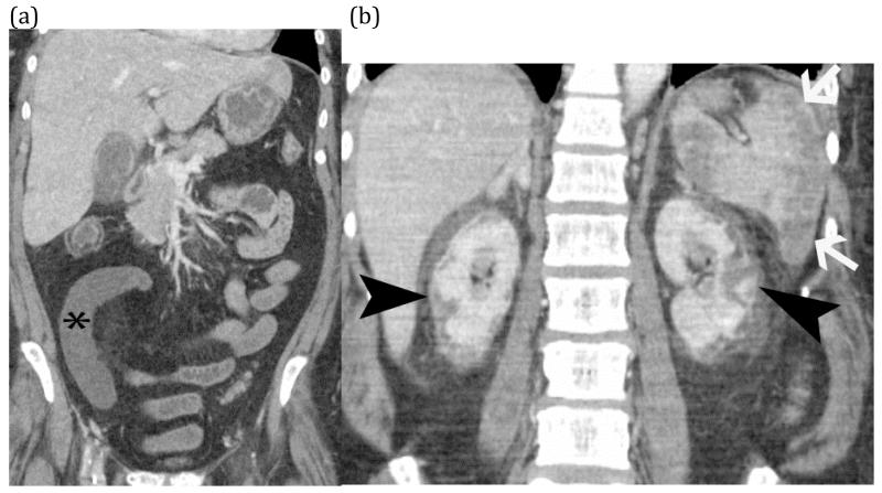
FIGURE 19 (a) and (b).
Shock bowel from hypotension – Coronal CT images of an elderly man with presented sepsis and hypotension. The first image (a) shows non-enhancement of a small bowel segment (*) compatible with small bowel ischemia. Additional images (b) shows evidence of global hypotension and shock with renal cortical necrosis (black arrow heads) and splenic infarcts (white arrows).

