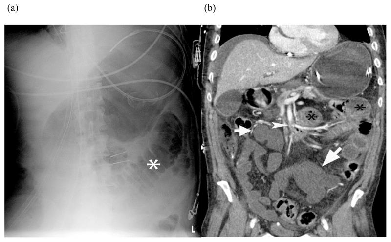
FIGURE 6.
Plain radiograph of small bowel ischemia – Plain radiograph (a) shows a focally dilated “paper thin” segment of small bowel (*) that had persisted over several consecutive examinations. Subsequent coronal reformatted CT image (b) of the same patient shows the corresponding dilated segment (*) as well as other fluid filled segments of small bowel with absent mural enhancement (white arrow) and clot in the SMA (white arrow head). At laparotomy, 220 centimeters of dead bowel was found.

