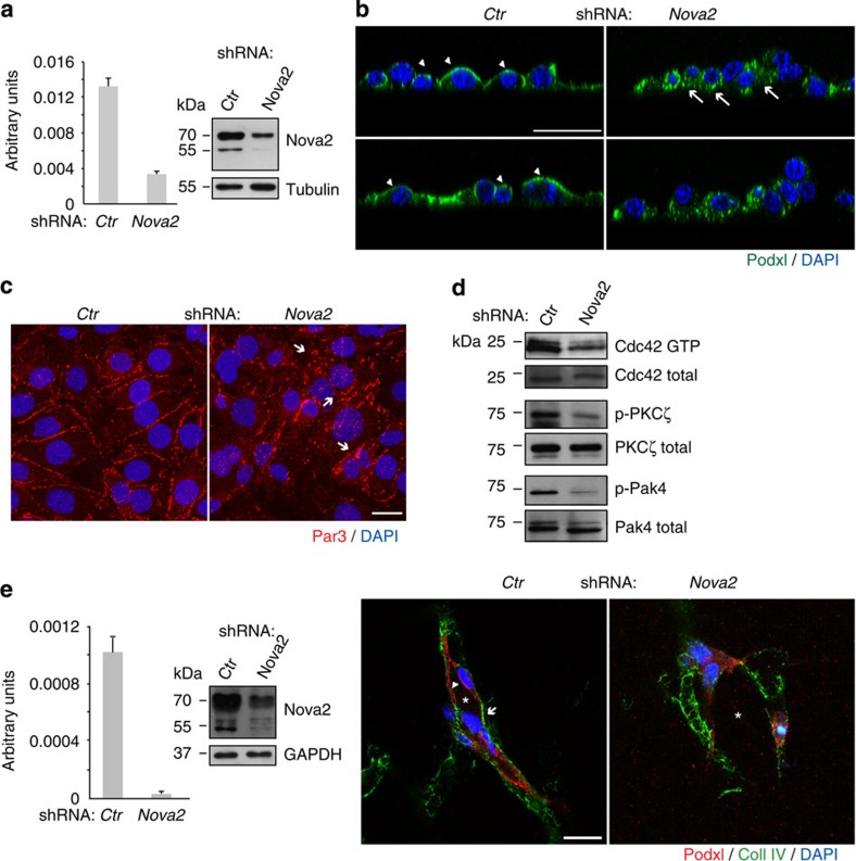
Figure 3. Nova2 is required for the EC polarization.
(a) Nova2 mRNA levels in knockdown mouse ECs (grown as confluent). The Nova2 protein level was analysed by immunoblotting using an anti-Nova2 antibody (Tubulin as loading control). (b) Immunofluorescence (IF) analysis of Podocalyxin (Podxl, green) and DAPI (blue) in 2D culture of control (Ctr) and Nova2-depleted ECs. Podxl is often distributed to the basal (arrows) instead of the apical surface (arrowheads) in Nova2-silenced ECs (confocal sections, z axis; scale bar, 25 μm). (c) IF analysis of Par3 (red) and DAPI (blue) in Ctr or Nova2 knockdown ECs. Arrows indicate altered and fragmented junctional staining of Par3 (bar 20 μm). (d) In vitro pull down of GTP-bound Cdc42 in Ctr and Nova2-silenced ECs. Immunoblotting for the phosphorylation status of PKCζ and Pak4 is also shown. (e) Left: Nova2 mRNA levels in knockdown HUVECs. The Nova2 protein level was analysed by immunoblotting using an anti-Nova2 antibody (GAPDH as the loading control). Right: in 3D collagen gel, control HUVECs form vascular structures with a central lumen (asterisk) and apical Podxl and basal collagen IV (Coll IV) proper localization (arrowheads and arrows, respectively), whereas Nova2-silenced HUVECs are not correctly polarized (scale bar, 20 μm). Error bars indicate mean ±s.d. calculated from three independent experiments (n=3).

