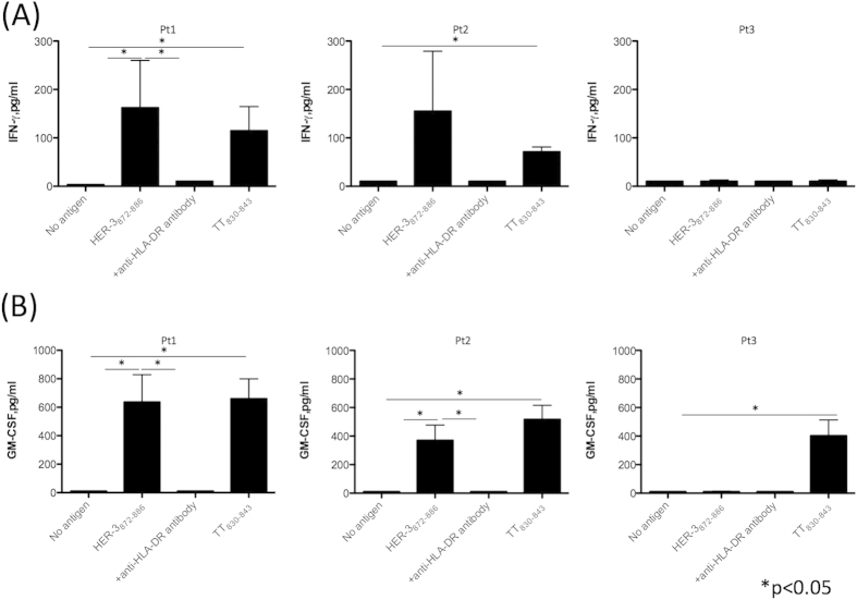
Figure 7. T cell responses to HER-3 peptide in PBMCs from HNSCC patients.
PBMCs from three HNSCC patients (Pt1: hypopharyngeal squamous cell carcinoma, Stage IV; Pt2: tongue squamous cell carcinoma, Stage IV, and Pt3: oropharyngeal squamous cell carcinoma, Stage IV) were stimulated with HER-3872−886 for seven days. Tetanus toxin830−843 was used as a positive control. After culture, PBMCs were re-stimulated with peptide-pulsed irradiated auto PBMCs for another seven days. Culture supernatant was collected to measure the production of IFN-γ (A) and GM-CSF (B). Columns: means of triplicate measurements, bars: SEM. The results shown are representative of 3 experiments that were performed on the same samples.

