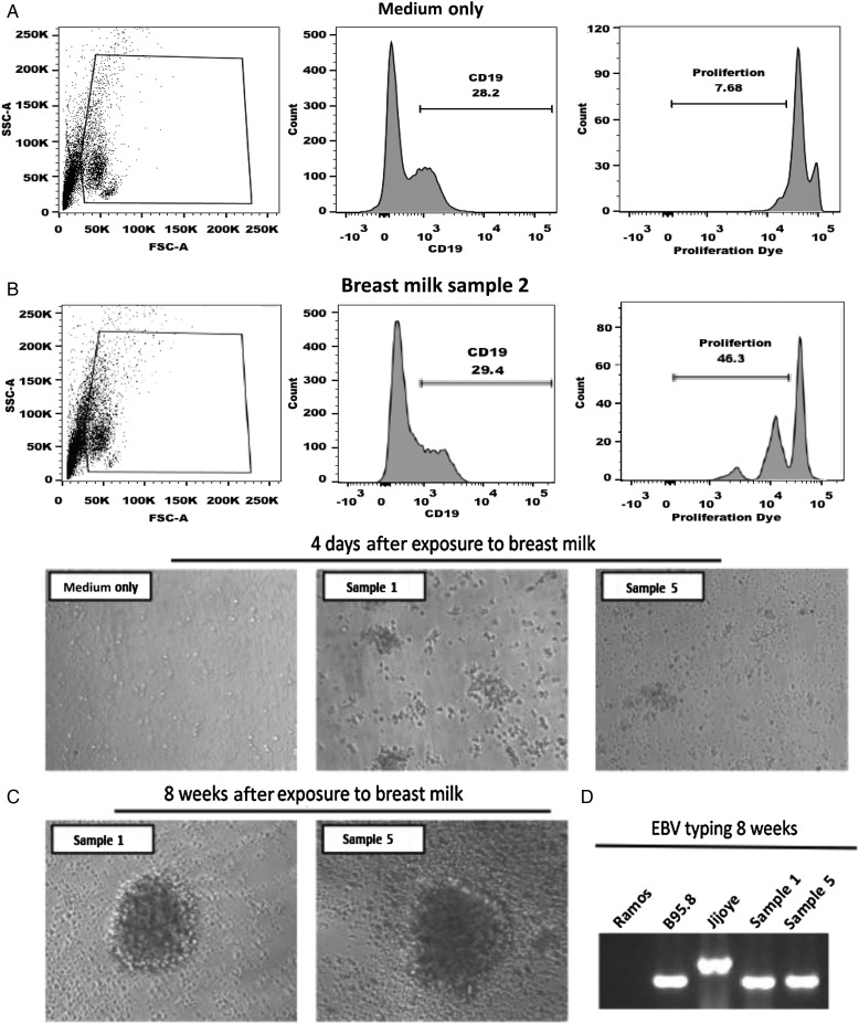
Figure 2.
Peripheral blood mononuclear cells (PBMCs) were labeled with e450 proliferation dye and exposed to breast milk collected at approximately postpartum week 6 or medium for 2 hours. Subsequently, cells were washed, cultured for 7 days, and analyzed for proliferation via flow cytometry. B cells were initially gated and analyzed for proliferation, as measured by diluting fluorescence intensity of e450 proliferation dye. A, Representative histograms of B-cell proliferation for 20 individual breast milk samples collected at approximately postpartum week 6; 2 PBMC donors per breast milk sample were tested. B, Prototypical Epstein-Barr virus (EBV)-induced cell aggregation was observed microscopically 4 days after exposure to breast milk. Representative images are shown. C and D, Following exposure to breast milk, PBMCs were cultured for 8 weeks and examined for generation of EBV lymphoblastoid cell lines. C, Microscopic images of 2 lymphoblastoid cell lines generated form PBMCs following exposure to breast milk collected at approximately postpartum week 6 are shown. D, Breast milk lymphoblastoid cell lines were analyzed by polymerase chain reaction (PCR) analysis of the EBNA3C encoding region to determine EBV type. The EBV-negative cell line Ramos served as a negative control, and B95.8 and Jijoye cell lines were used as positive controls for type 1 and type 2 EBV, respectively. PCR product sizes were as expected for EBV type 1 and type 2.

