Figure 1.
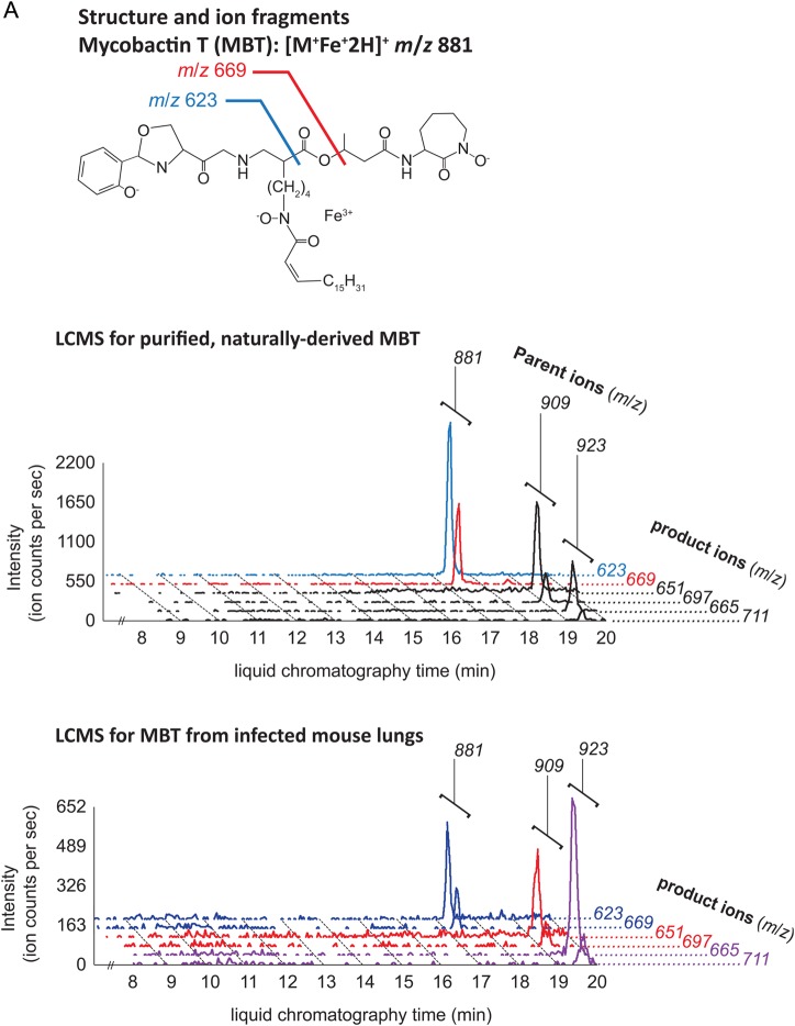
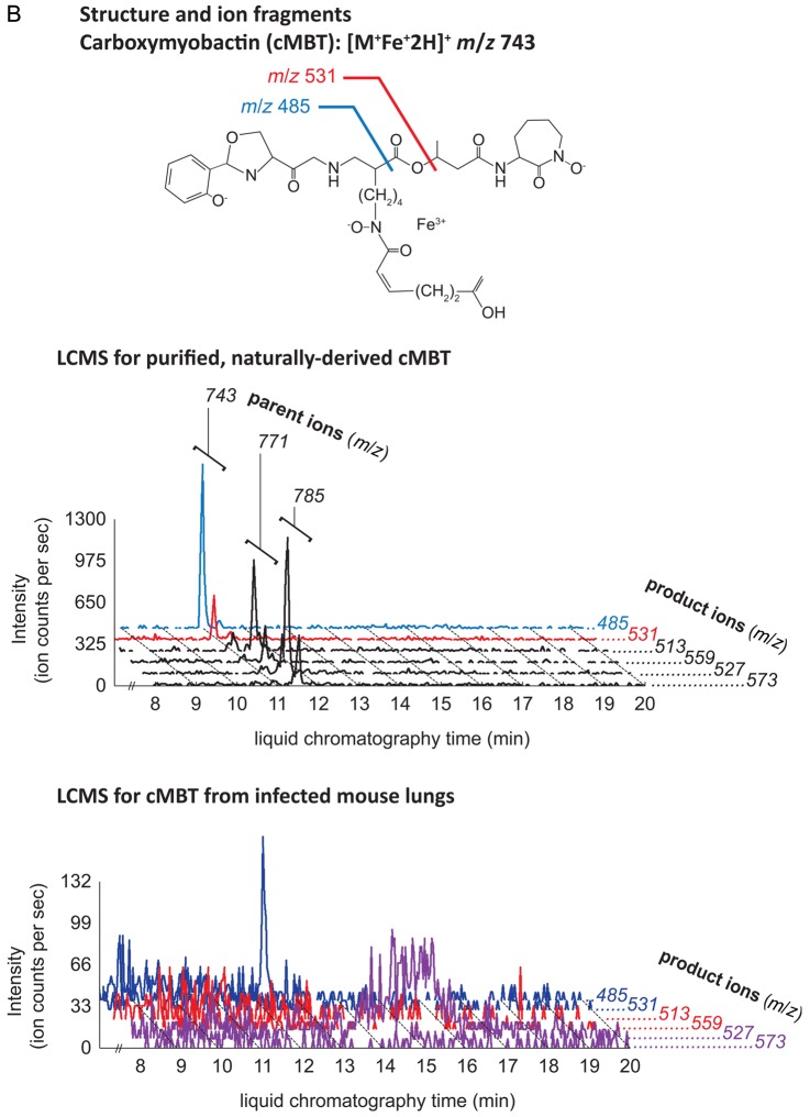
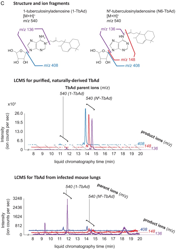
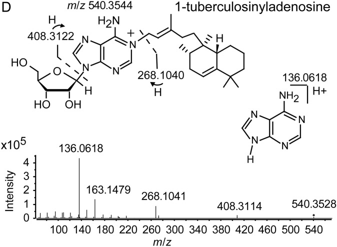
Structures and ion fragments (top), representative ion chromatograms of purified, naturally derived, tuberculosis-specific bacterial small molecules (BSMs), illustrating the characteristic ion intensity peaks with collision-induced liquid chromatography mass spectrometry (LCMS) in positive ion mode at concentrations of 10 nM (middle), and characteristic ion chromatograms of the product ions detected by LCMS analysis from infected mouse lungs (bottom) for mycobactin T (A), carboxymycobactin (B), and tuberculosinyladenosine in its 2 forms, 1-TbAd and the isomeric N6-TbAd (C). For mycobactin T, the 15 methylene aliphatic side chain as shown gives a parent ion m/z of 881, but 17- (m/z 909) and 18-methylene (m/z 923) forms are also produced. For carboxymycobactin, the aliphatic link as shown gives a parent ion m/z of 743 but the 2-methylene aliphatic chain shown may extend to 4 (m/z 771) or 5 methylenes (m/z 785) in length. For mouse infections, LCMS analyses are shown from 1 processed lung tissue sample from a BALB/c mouse sacrificed 21 days after aerosol infection with M. tuberculosis H37Rv. Infected mouse lungs showed characteristic peaks for mycobactin T (A, bottom panel) and 1-TbAd (C, bottom panel), but carboxymycobactin was less abundant with only 1 ion transition observed above the detection limit (B, bottom panel). Trace levels of N6-TbAd were observed eluting later than 1-TbAd in the infected mouse lung (C, bottom panel). The 1-TbAd used as standard material in this study was fully characterized by mass spectrometry and nuclear magnetic resonance (NMR) spectroscopy. This included accurate mass assignments (elemental composition), collisionally induced dissociation fragment analysis, and 2-dimensional NMR, allowing characterization of the 1-TbAd structure (D). Abbreviation: TbAd, tuberculosinyladenosine.


