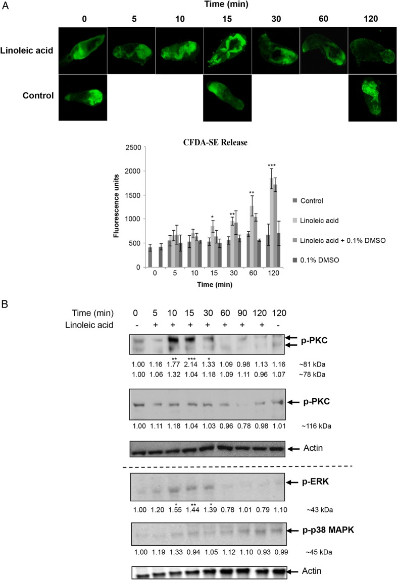
Figure 4.
Effect of linoleic acid (LA) on release of acetabular gland components and protein kinase C (PKC), extracellular signal–regulated kinase (ERK), and p38 mitogen-activated protein kinase (p38 MAPK) signaling in Schistosoma mansoni cercariae. A, Release of cercariae acetabular gland components (labeled with carboxyfluorescein diacetate succinimidyl ester [CFDA-SE]; green) following LA exposure. Images are representative of cercariae populations obtained from 2 independent experiments. Cercariae preincubated with CFDA-SE were or were not exposed to LA, with or without 0.1% dimethyl sulfoxide (DMSO), and mean fluorescence (±SD) was determined over 120 minutes (bottom; n = 3). *P ≤ .05, **P ≤ .01, and ***P ≤ .001, by analysis of variance (ANOVA), when compared to control or 0.1% DMSO values. B, Cercariae were (for 5–120 minutes) or were not (data are from 0 and 120 minutes; controls) exposed to LA, and proteins were processed for Western blotting with anti-phospho PKC (ζ Thr410), anti-phospho PKC (βII Ser660), anti-phospho p44/42 MAPK (ERK), anti-phospho p38 MAPK, and anti-actin antibodies. Blots from at least 3 independent experiments were analyzed for band intensity, and intensity values were normalized for actin. Mean change in phosphorylation (shown under each blot) was determined relative to 0-minute controls, which were assigned a value of 1. *P ≤ .05, **P ≤ .01, and ***P ≤ .001, by ANOVA. This figure is available in black and white in print and in color online.

