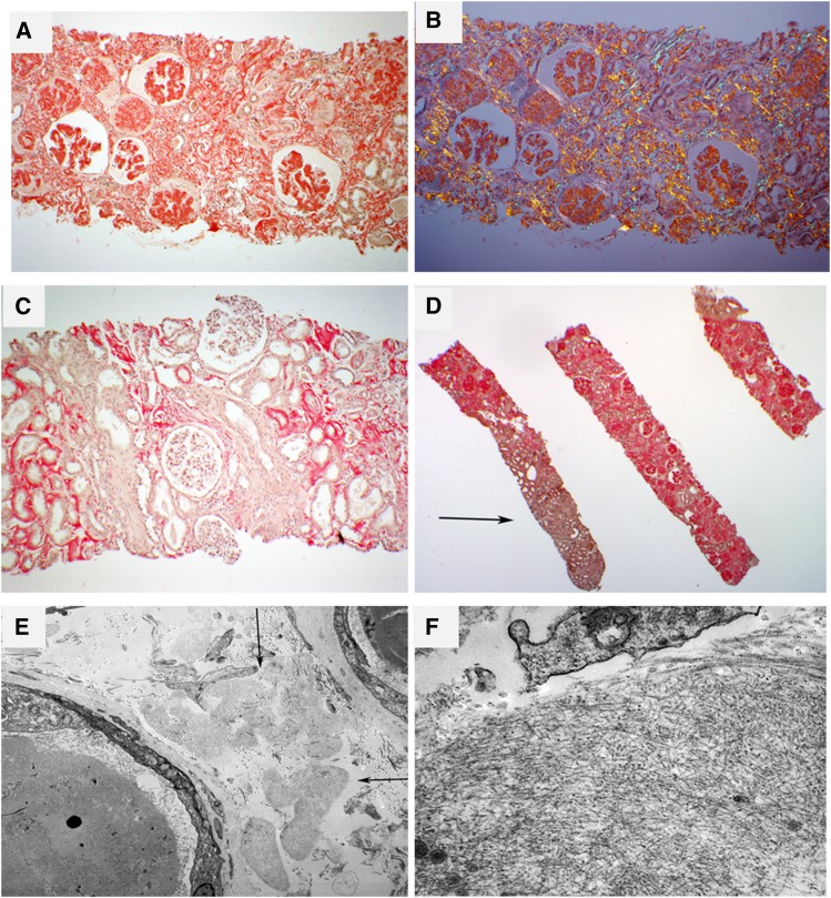
Figure 2.
Renal pathology in amyloidosis derived from leukocyte cell–derived chemotaxin 2 (ALECT2) amyloidosis. (A) The case depicted shows extensive interstitial, glomerular, and vascular congophilic amyloid deposition. This patient had subnephrotic proteinuria (2.5 g/d) at diagnosis. Magnification, ×40. (B) Same field as in A. The congophilic amyloid deposits show anomalous colors (red, yellow, and green) under polarized light. Magnification, ×40. (C) The case depicted is from a patient who had a serum creatinine of 2.3 mg/dl at biopsy without proteinuria. There is diffuse cortical interstitial and focal arteriolar amyloid deposition with sparing of glomeruli (Congo red stain). Magnification, ×100. (D) This patient exhibits extensive involvement of cortical interstitium and glomeruli. The medullary interstitium (arrow) is spared (Congo red stain). Magnification, ×20. (E) A low-power electron microscopic image showing several interstitial collections of amyloid deposits (arrows). Magnification, ×5800. (F) A higher-magnification image reveals the fibrillar substructure of deposits. Magnification, ×49,000.

