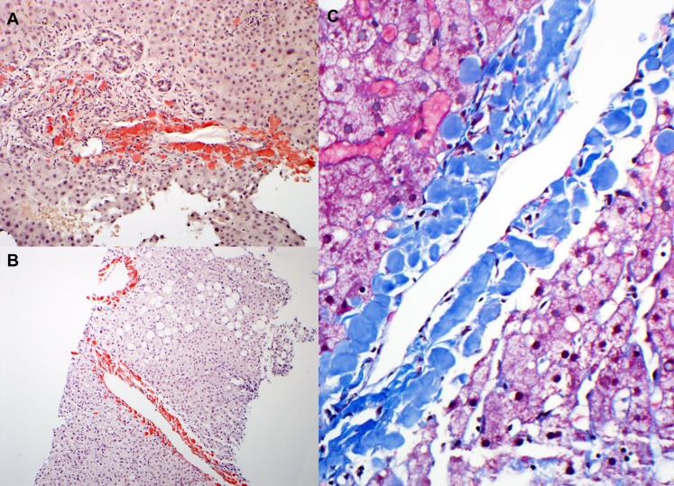
Figure 3.
Liver pathology in amyloidosis derived from leukocyte cell–derived chemotaxin 2 (ALECT2) amyloidosis. (A) There are periportal deposits of strongly congophilic amyloid deposits. Magnification, ×100. (B) In this low-power image, strongly congophilic amyloid deposits are seen surrounding the central veins. In contrast to Ig light chain–derived amyloidosis, the perisinusoidal spaces are spared. Magnification, ×40. (C) On high magnification, ALECT2 deposits form large acellular globules, which appear blue on trichrome stain. Magnification, ×200.

