Fig. 6.
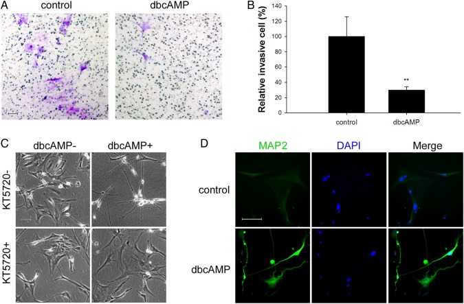
Representative effects of dbcAMP on human glioblastoma (GBM) primary cultures. Human GBM samples were collected soon after surgery and immediately cultured. (A and B) Transwell assays were performed with primary GBM cells after 24 hours of treatment with/without dbcAMP (1 mM) as indicated. About 5 × 10⁴ viable cells were seeded into the upper chamber. Invasive cells were counted and normalized with number of invasive cells of the control group. The random representative fields of an experiment are shown in panel A (original magnification: × 100; scale bar: 100 μm), and the corresponding statistics are shown in panel B (data are shown as the mean ± SD (n = 3). **P < .01. The experiments were repeated at least 3 times before statistical analysis. (C) Cells were seeded at a density of 5 × 10⁴and pretreated with/without 2 μM PKA inhibitor KT-5720 for 2 hours followed with/without 48 hours of treatment of 1 mM dbcAMP. Phase contrast microscopy evaluated the morphological changes (original magnification: × 100; scale bar: 100 μm). (D) Primary GBM cells were seeded at a density of 5 × 10⁴, treated with/without 1 mM dbcAMP for 48 hours, and an immunofluorescence assay was performed to examine the expression of MAP2 (original magnification: × 200; scale bar: 100 μm).

